左肺尖Pancoast瘤(肺上沟癌)
History: A 45-year-old woman presents with pain in her upper back and persistent pain and limited use of her left arm.
医学百科网 | YxBaike.Com
病史:45岁女性,上背部疼痛,左臂持续疼痛,运动受限。 医学百科网 | YxBaike.Com
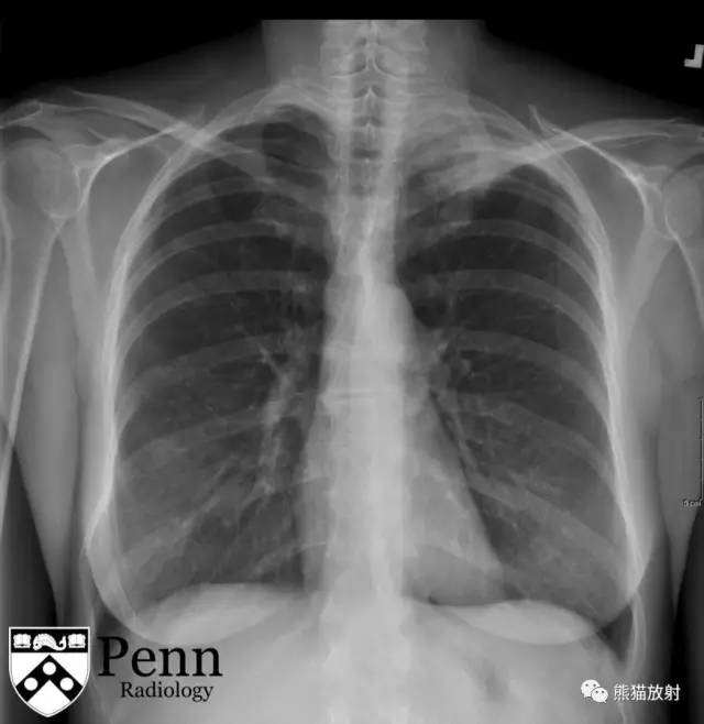
Posteroanterior (PA) radiograph of the chest is shown below.
胸部正位片如下所示。 医学百科网 | YxBaike.Com
 医学百科网 | YxBaike.Com
医学百科网 | YxBaike.Com
CT images 医学百科网 | YxBaike.Com
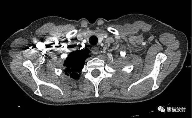
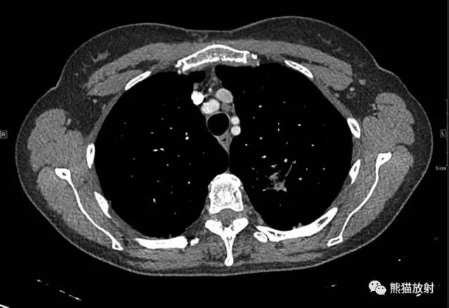
Axial contrast-enhanced CT images of the thorax in soft-tissue windows are shown below. 医学百科网 | YxBaike.Com
胸部CT增强扫描(软组织窗)如下所示。
医学百科网 | YxBaike.Com

Additional CT images
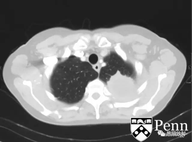
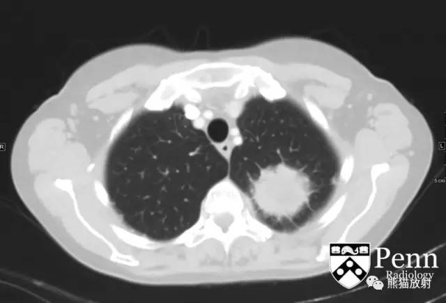
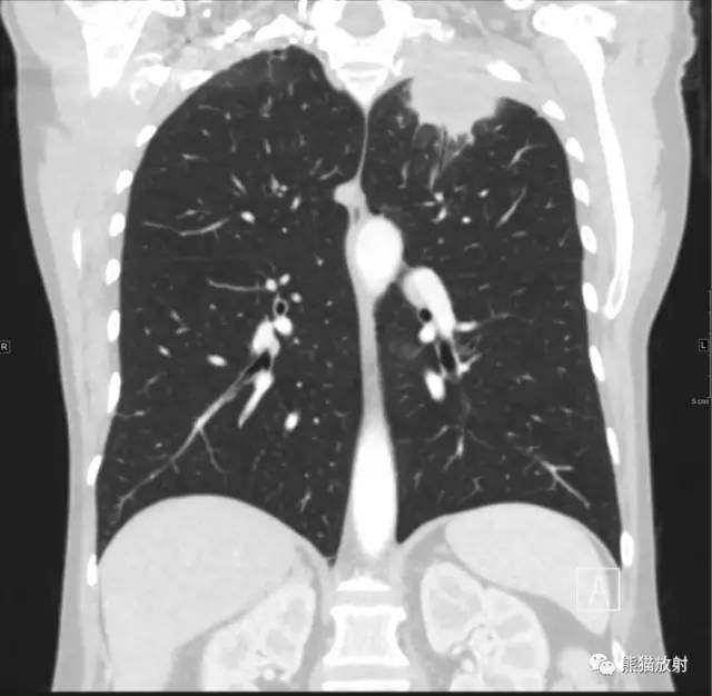
Axial and coronal contrast-enhanced CT images of the thorax in lung windows are shown below.
胸部CT增强轴位及冠状位肺窗图像如下所示。 医学百科网 | YxBaike.Com
医学百科网 | YxBaike.Com
Findings
医学百科网 | YxBaike.Com
- Chest radiograph: The PA radiograph of the chest reveals an opacity in the left upper lung apex.
- Chest CT: Contrast-enhanced CT of the chest revealed a centrally necrotic spiculated mass, abutting the pleura measuring 4.7 x 4.9 x 3.2 cm. There may be extra pleural extension; however, no definite osseous destruction is visualized. There is a 14 x 10 mm left supraclavicular lymph node (image 3) and a left suprahilar node that measures 19 x 11 mm (image 5). There is no contralateral mediastinal or right-sided hilar adenopathy. These findings are within a background of apical-predominant paraseptal emphysema.
影像表现:
医学百科网 | YxBaike.Com
- 胸片:胸部正位片示左肺尖阴影。
- 胸部CT扫描:增强扫描示左肺尖胸膜下肿物,大小约4.7 x 4.9 x 3.2 cm,中心坏死,周围可见毛刺;病变侵犯至胸膜外,未见明显骨质破坏。左锁骨上及左上肺门分别见14 x 10 mm、19 x 11 mm增大淋巴结。对侧纵隔及右肺门未见肿大淋巴结。肺尖可见多发小叶间隔旁肺气肿。
Differential diagnosis 医学百科网 | YxBaike.Com
- Pancoast tumor
- Pulmonary metastasis
- Pleural metastasis
- Mesothelioma
- Lymphoma
- Tuberculosis
- Nocardiosis
- Actinomycosis
鉴别诊断: 医学百科网 | YxBaike.Com
- 肺上沟癌
- 肺转移瘤
- 胸膜转移
- 间皮瘤
- 淋巴瘤
- 结核
- 诺卡氏菌病
- 放射菌病
Diagnosis: Pancoast tumor — primary bronchogenic carcinoma 医学百科网 | YxBaike.Com
诊断:Pancoast瘤(原发性支气管肺癌) 医学百科网 | YxBaike.Com
Discussion
Pancoast tumors
Pathophysiology 医学百科网 | YxBaike.Com
Pancoast tumors are also referred to as superior sulcal tumors. This term is often reserved for bronchogenic carcinomas. The name is derived from their association with Pancoast syndrome, which results when the tumor involves the brachial plexus and sympathetic chain. Pancoast syndrome consists of shoulder pain, Horner syndrome, and C8-T2 radicular pain. 医学百科网 | YxBaike.Com
病理生理学:Pancoast瘤,也称作肺上钩癌,一般为支气管肺癌。因Pancoast综合征而得名,肿瘤累及臂丛神经及交感链。Pancoast综合征包括:肩部疼痛、霍纳综合征、C8-T2神经根痛。
Epidemiology
医学百科网 | YxBaike.Com
Pancoast tumors account for 3% to 5% of all bronchogenic carcinomas. Most affected patients are between 50 and 60 years. Men are more commonly affected than women. Risk factors include smoking, asbestos exposure, and exposure to industrial elements.
流行病学:Pancoast瘤占所有支气管肺癌的3-5%,好发年龄位于50-60岁,男性多于女性。危险因素包括吸烟、石棉接触、暴露于工业环境。 医学百科网 | YxBaike.Com
Clinical presentation 医学百科网 | YxBaike.Com
Patients present with symptoms of Pancoast syndrome: shoulder pain, Horner syndrome, and C8-T2 radicular pain. Patients may also have upper arm edema, secondary to subclavian vein occlusion. 医学百科网 | YxBaike.Com
临床表现:患者主要表现为Pancoast综合征:肩部疼痛、霍纳综合征、C8-T2神经根痛。也可因锁骨下静脉闭塞表现为上臂水肿。 医学百科网 | YxBaike.Com
Imaging features
医学百科网 | YxBaike.Com
- Plain radiograph: Plain films reveal an opacity at the apex of the lungs in approximately two-thirds of cases. Some films will reveal an asymmetric unilateral apical cap. There can be rib involvement or extension into the supraclavicular fossa.
- CT: Nonenhanced CT can be used to confirm the presence of a mass. Additionally, it can be used to assess skeletal involvement. Metastatic disease to the lungs, pleura, chest wall, and upper abdomen can be limitedly evaluated via noncontrast scan. Contrast-enhanced CT is useful to evaluate vascular involvement and node size/involvement.
- PET/CT: Uptake on PET/CT can be used to evaluate the primary neoplasm, lymph node metastases, and distant metastases.
- MRI: MRI is useful to evaluate the soft-tissue involvement of Pancoast involvement, especially the brachial plexus.
影像表现: 医学百科网 | YxBaike.Com
- 平片:约2/3的病例表现为肺尖阴影,部分表现为一侧肺尖形态不规则,可见肋骨受累,病变上凸至锁骨上窝。
- CT:平扫可明确有无肿物,骨质结构有无受累,对于评价肺内、胸膜、胸壁、上腹部有无转移有一定限度。增强扫描可评估血管受累及淋巴结转移情况。
- PET/CT:原发肿瘤、淋巴结转移及远处转移灶有摄取。
- MRI:可用于评估臂丛神经有无受累。
Treatment 医学百科网 | YxBaike.Com
Treatment of Pancoast tumor is largely dependent on the stage of the cancer and the involvement of the apex, including soft-tissue structures such as nerves and vessels. If there is involvement of the brachial plexus and subclavian vessels, chemoradiation is often used to down grade the tumor to increase the probability of resection. 医学百科网 | YxBaike.Com
治疗: 医学百科网 | YxBaike.Com
Pancoast瘤的治疗主要根据肿瘤的分期及局部受累(血管、神经)的情况而定。如果肿瘤侵犯臂丛神经及锁骨下血管,通常借助预先的放化疗来降低肿瘤分级来增加可切除率。
医学百科网 | YxBaike.Com
附件列表
词条内容仅供参考,如果您需要解决具体问题,请遵医嘱或咨询医师。
本站词条内容如果涉嫌侵权,请与客服联系,我们将及时进行处理。如需转载,请注明来源于www.yxbaike.com
如果您认为本词条还有待完善,请 编辑
上一篇 “桃尖征” 的影像表现与临床意义 下一篇 右肺下叶背段周围性肺癌1例CT
