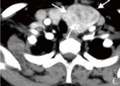
Findings
Differential diagnosis
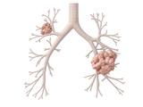
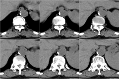
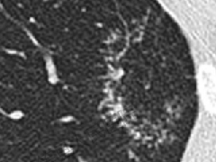
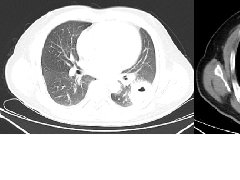
Diagnosis: Pulmonary alveolar proteinosis
The patient underwent bronchoalveolar lavage, which demonstrated milky and turbid fluid with thick sediment. Cytology demonstrated large foamy macrophages. Serum antigranulocyte-macrophage colony-stimulating factor (anti-GM-CSF) autoantibodies were positive.

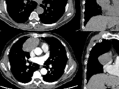
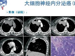 纵隔大细胞神经内分泌癌1例CT影像
 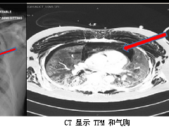 张力性纵隔气肿影像表现及严重度分级
  迅速增大的肺部结节,首先考虑良性,确诊需要肺穿
 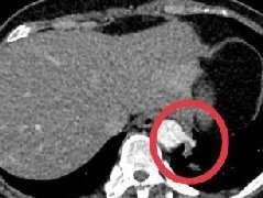 肺隔离症:易误诊为肺癌的占位性病变,肺穿刺禁忌!
  肺段与肺内管道应用解剖
  肺转移瘤的十种不典型CT表现
  肺内淋巴结的CT表现特点及与病理对照
  肺实变与肺不张的CT鉴别诊断
  医生现身说法,这五种忙帮不得!
  北大教授:要真正让医务人员有阳光体面的收入!医
  为值夜班的医生护士鼓与呼:请给我们更多关注!
  广东拟取消医院用药数量限制,满足患者多样性需求
  博士、硕士入职就给精装房!又有医院不惜下血本招
  历时7年之久,温医生宣判无罪!
  重磅!四川发文:严禁限制医生多点执业
  与真人医生诊断一致性达96%:AI医生应用前景广阔
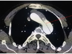
辽ICP备15019299号-3